PYDL Yeast Display Vector Series
Next-Generation Yeast Display Vectors for Antibody and Protein Engineering.
The PYDL Vector Series is a line of plasmids dedicated to Yeast Display, a technology renowned for revolutionizing protein engineering and antibody discovery. Leveraging our expertise in molecular biology, these vectors provide a robust and versatile platform for displaying diverse protein libraries on the surface of Saccharomyces cerevisiae cells, enabling rapid screening and selection of variants with desired properties.
Overview of Yeast Display Technology
Yeast display is a powerful method where engineered proteins are presented on the surface of S. cerevisiae cells, allowing for high-throughput screening.
Key Advantages
-
Eukaryotic Expression: Proteins are folded and modified in a native-like eukaryotic environment, increasing the chance of functional display for complex proteins like antibodies or receptors.
-
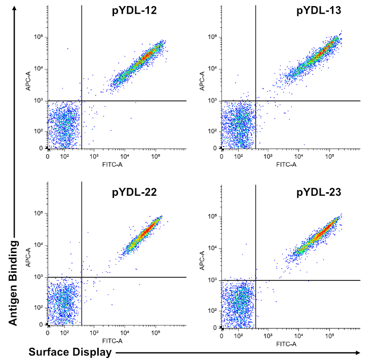
Quantitative Analysis: Flow cytometry enables highly sensitive, quantitative, and high-throughput screening of library members, facilitating the isolation of rare high-affinity binders.
-
Facile Genetic Manipulation: Yeast is genetically tractable, which simplifies library construction and mutagenesis.
-
Direct Linkage: The displayed protein is genetically linked to its encoding DNA, simplifying the recovery and sequencing of selected clone.
-
Versatility: The system is suitable for displaying a wide range of proteins, including scFvs and VHHs (single-domain antibodies).
How it Works
The system utilizes the native mating agglutination machinery, specifically the Aga1p/Aga2p components of S. cerevisiae.
-
The protein of interest (POI) is expressed as a fusion protein with Aga2p in yeast strains (like EBY100) that also express Aga1p.
-
The Aga2p-POI fusion forms two critical disulfide bonds with the larger, cell-wall-anchored component, Aga1p.
-
This covalent linkage creates a stable, display complex that anchors the POI to the yeast cell surface, making it accessible for directed evolution, binding assays, and flow cytometry screening.
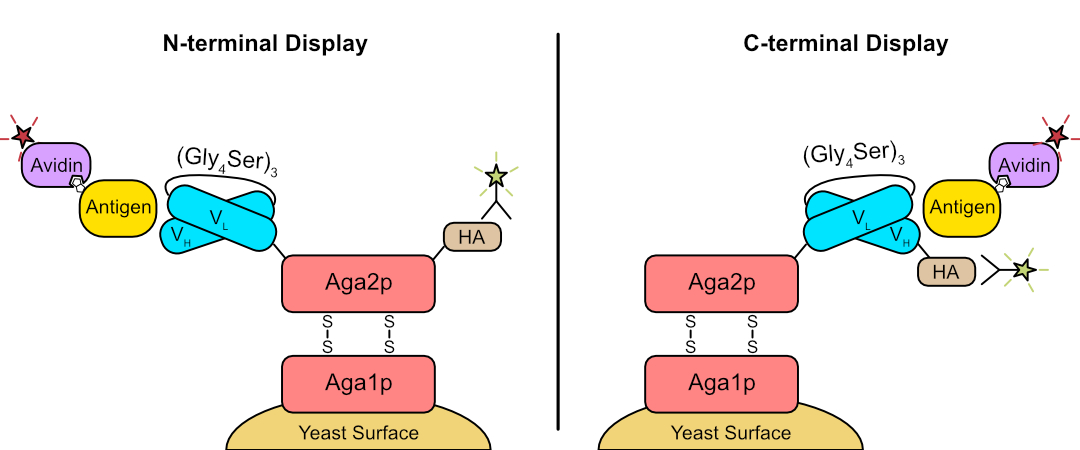
Figure 1. Yeast surface display. Representation of N- and C-terminal Aga2p fusions for display of an scFv (as an example) on yeast. Surface display is detected by antibody staining of epitope tag (HA, or similar) and target binding using biotinylated antigen followed by fluorescently-labeled streptavidin.