Cart is empty
Catalog
Recently Viewed
CM13d3 - pIII-Defective Helper Phage
Technical Description
CM13d3 is a helper phage engineered for multivalent phage display. CM13d3 is a derivative of M13KO7 (1) having a wild-type pIII phenotype which allows efficient bacterial transduction but lacking a functional pIII gene thus driving the display of pIII-fusion proteins expressed by the phagemid on the phage head (2). The use of pIII-defective helper phage has been shown to increase display of scFv by a factor up to 100-fold and more (3-5). This preparation contains enough helper phage to superinfect up to 0.5 L (500 ml) of TG1 culture. Other bacterial strains may require different amounts of CM13d3 helper phage. More information can be found here.
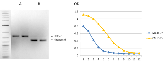
Figure 1. CM13d3 Packaging & Display. Virions derived from pADL-22c phagemid displaying HyHEL-10 scFv were prepared with CM13d3 helper phage. Single stranded DNA analysis of the virion DNA was analyzed by gel electrophoresis (left panel). Incorporation of phagemid DNA is exquisite with very little helper ssDNA (B). The helper ssDNA alone is shown on the left (A). The DNA ladder in made of double stranded DNA ans sizes are irrelevant. Analysis of virion binding to lyzozyme by phage ELISA shows a 10-fold binding improvement comparatively to the same virions prepared with the non-defective M13KO7 helper (right panel).
Applications
-
Phage display.
For research use only; not intended for any animal or human therapeutic or diagnostic use.
Source
CM13d3 virions were isolated from the supernatant of infected E. coli TG1 cells with a functional gene III and purified by PEG precipitation.
Specifications
Composition: 50% glycerol TBS buffered.
Concentration: >2.0 x10¹¹ cfu/ml.
Storage temperature: -20°C.
Product size: 0.5 ml.
Quality Control & Certification of Analysis
Virion concentration
Infectivity is measured as cfu after transduction of TG1 cells. Pfu counts on pIII-supplemented bacteria are ~4 times higher.
References
- Vieira, J. and Messing, J. (1987) R. Wu and L. Grossman (Eds.), Methods Enzymol., 153, pp. 3-11. San Diego: Academic Press.
- Dueñas, M. AND Borrebaeck CA. (1995). Novel helper phage design: intergenic region affects the assembly of bacteriophages and the size of antibody libraries, FEMS Microbiol lett., 125(2-3):317-21.
- Rondot, S., Koch, J., Breitling, F. and Dübel, S. (2001). A helper phage to improve single-chain antibody presentation in phage display. Nat Biotechnol., 19(1):75-8.
- Broders O, Breitling F, Dübel S (2003). Hyperphage – Improving antibody presentation in phage display, Methods Mol Biol, 205, 295-302.
- Soltes G, Hust M, Ng KKY, Bansal A, Field J, Stewart DIH, Dübel S, Cha S, Wiersma EJ (2007). On the influence of vector design on antibody phage display, J Biotechnol. 127, 626-637.
Related Products
Citations
-
Hampton JT, Lalonde TJ, Tharp JM et al. (2022). Novel Regioselective Approach to Cyclize Phage-Displayed Peptides in Combination with Epitope-Directed Selection to Identify a Potent Neutralizing Macrocyclic Peptide for SARS-CoV-2. ACS Chem. Biol., 17(10):2911-2922.
-
Jester BW, Zhao H, Gewe M et al. (2022). Development of spirulina for the manufacture and oral delivery of protein therapeutics. Nat Biotechnol., Mar 21.