Cart is empty
Catalog
Recently Viewed
CM13K - Trypsin-Sensitive Helper Phage
Technical Description
CM13Kis a helper phage especially engineered for phage display. CM13K is a derivative of CM13 (cat# PH020L) that contains a trypsin-sensitive linker between the N2 domain and the CT domain of the minor coat protein III. CM13K is therefore very similar to KM13 helper phage (1) and AGM13 helper phage (2). Upon exposure to trypsin, the N1N2 domains are excised and CM13K protein III becomes unable to mediate transduction. Trypsinization of the CM13K itself results in a decrease of the number of pfu/ml by 1 x 10⁶. Therefore, transduction after trypsin treatment will almost exclusively derive from fusion phage bearing a wild-type, trypsin-resistant protein III typically made by the phagemid (Fig. 1). Moreover, it has been shown that trypsin release of bound phage is far superior to classical acidic elution (3). Together with the inactivation of wild type protein III, the use of CM13K and trypsin elution results in more efficient and more selective biopannings.
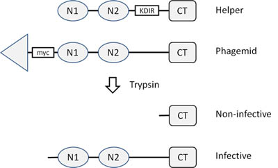
Figure 1. CM13K and trypsin elution. CM13K has a trypsin site between the N2 domain and the CT (C-terminal) domain of pIII. After treatment by trypsin, the N1 and N2 domains are removed and the helper phage protein III is inactivated. Phage bearing a fusion protein on pIII and a trypsin-sensitive tag (e.g. Myc tag) are eluted and remains active.
Applications
-
Phage display.
For research use only; not intended for any animal or human therapeutic or diagnostic use.
Source
CM13K virions were isolated from the supernatant of infected E. coli TG1 cells and purified by PEG precipitation.
Specifications
Composition: 50% glycerol TBS buffered.
Concentration: 2 x10¹² pfu/ml; 1 x10¹³ virions/ml; infectivity ~20%
Storage temperature: -20°C.
Product size: 5 x 1 ml.
Quality Control & Certification of Analysis
Virion concentration:
Phage DNA concentrations are determined by UV spectrophotometry and virion concentrations calculated based on the length of CM13K genome.
Phage titer:
CM13K preparations are serially diluted and mixed with melted LB top agar and TG1 cells. The mixtures are poured over LB bottom agar plates and incubated overnight at 37°C; the morning after plaques are counted. Controls include plates without virions and have no visible plaques.
Infectivity:
Infectivities are counted as the ratio between phage titer and virion concentration. Infectivities between 15 and 20% are expected.
Contamination check:
Phage preparations show no growth on 2xYT agar plates.
Certification:
Products meet all specifications.
Notes
Conditions for optimal transduction:
Always use bacteria freshly made from the night before; typically grow your phagemid-containing bacteria overnight in 2xYT medium from a single colony in the presence of ampicillin and glucose at 30°C or 37°C. Dilute the bacterial culture 1:20 v/v with fresh 2xYT medium and incubate for one hour at 37°C. Measure the absorbance at 600 nm, at best using a large 1 ml cuvette and a 1:5 or a 1:10 dilution; adjust to 0.5 OD and add CM13 between 1 x 10(9) and 1 x 10(10) pfu/ml. We recommend 2 x 10(9) pfu/ml or 1 µl of CM13 preparation per ml of culture as a good balance between not using too much helper and achieving a high level of superinfection; more helper may yield more phage but results are often inconsistent.
Immediately transfer the culture to a shaker and incubate for 30 min to 1 h at 37°C and 250 rpm; preincubation of helper and bacteria on the bench or at 37°C without shaking is unnecessary and often leads to a greater variability in phage yields; best superinfections are obtained at 37°C and 250 rpm. Then add kanamycin 50 µM, ampicillin 100 µM and IPTG 200 µM if you are using pADL-10b, pAK100 or pAK200 phagemid and lower the temperature to 30°C; harvest the phage after 8 h to o/n.
Influence of cell density:
An F+ bacterial strain containing the F plasmid is required for superinfection by CM13 virions. Removal of glucose, induction of the Lac promoter and expressing the fusion coat protein at possible toxic levels may cause the bacteria to stop growing; therefore, it is better to add the helper phage at the highest cell density possible. Inversely the presence of a large percentage of non-transduced bacteria can lead to low phage production; this problem is accentuated by a natural immunity to M13 superinfection caused by endogenous expression of full-length protein III, including expression by the phagemid. We have determined that the optimal cell density at which the helper phage is added to maximize phage production is at an optical density between 0.4 and 0.5 at 600 nm for TG1 and SS320 cells. In these conditions, use of a truncated protein III to limit immunity to superinfection is not necessary.
References
- Kristensen P, Winter G. Proteolytic selection for protein folding using filamentous bacteriophages. Fold Des. 1998;3(5):321‐328.
- Gupta A, Shrivastava N, Grover P, et al. A novel helper phage enabling construction of genome-scale ORF-enriched phage display libraries. PLoS One. 2013;8(9):e75212.
- Thomas WD, Smith GP. The case for trypsin release of affinity-selected phages. Biotechniques. 2010;49(3):651‐654.
Related Products
Citations
-
Mendoza-Salazar I., Gómez-Castellano KM., González-González E., Gamboa-Suasnavart R., Rodríguez-Luna SD., Santiago-Casas G., Cortés-Paniagua MI., Pérez-Tapia SM., Almagro JC. (2022). Anti-SARS-CoV-2 Omicron Antibodies Isolated from a SARS-CoV-2 Delta Semi-Immune Phage Display Library. Antibodies (Basel),10;11(1):13.